

Endometrioma, often referred to as a “chocolate cyst,” is a gynecological condition that affects women of reproductive age. With the help of imaging methods (US and MRI), doctors can identify and diagnose endometrioma with greater accuracy. This article demonstrates the role of imaging methods in the evaluation of endometrioma.
What is Endometrioma?
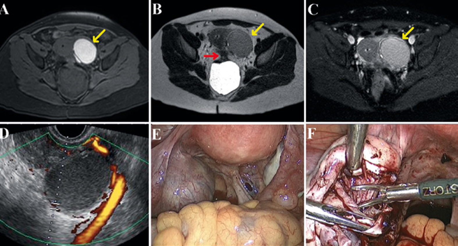
Endometrioma is a subtype of endometriosis, a condition where tissue similar to the uterus lining grows outside the uterus. Endometrioma specifically refers to cysts formed by this ectopic endometrial tissue, predominantly found on the ovaries. These cysts are filled with old, dark blood, giving them a brownish color and the nickname ‘chocolate cysts.’ The condition can lead to severe pelvic pain, infertility, and decreased ovarian function, impacting a woman’s quality of life significantly.
Importance of ultrasound (US) and Magnetic Resonance Imaging in the Diagnosis of Endometrioma
Ultrasound is a non-invasive imaging technique that uses sound waves to produce images of the body’s internal structures. It is an initial tool in the diagnosis of endometrioma, presenting high sensitivity and specificity for the diagnosis, allowing visualization of the cyst and evaluation of its characteristics.
Magnetic Resonance Imaging has sensitivity and specificity similar to ultrasound for endometriomas larger than 1 cm, but its sensitivity becomes higher when compared to US for small endometriomas smaller than 1 cm. Furthermore, MRI demonstrates the characteristics of the internal content of the evaluated cyst, being indicated when there are diagnostic doubts on US, enabling the differentiation of other diagnoses such as mucinous cystadenoma, teratomas, hemorrhagic cyst and lesions with risks of malignant neoplasia.
Differentiating endometrioma from other types of ovarian cysts is essential for defining the appropriate treatment plan.
Identifying Endometrioma via Ultrasound
Endometriomas have distinct ultrasound features that differentiate them from other ovarian cysts. They typically appear as cysts with diffuse, low-level echoes, often referred to as “ground glass” echogenicity. This characteristic is due to the thick, dark liquid inside the cyst. These can be bilateral in up to 40 to 50% of cases, present liquid-liquid levels, and can be uni or multiloculated, making it possible to identify the presence of peripheral clots, echogenic foci on their surface and hypo or avascular on Doppler.
Identifying endometrioma via Magnetic Resonance Imaging
On MRI, endometriomas can represent a typical pattern characterized by hypersignal on T1 in sequences with and without fat saturation, intermediate signal on T2 (shading sign), this due to the predominance of methemoglobin as a hemoglobin degradation product. T2 dark spot sign can also be identified due to chronic intralesional hemorrhage.
Association between endometrioma and deep pelvic endometriosis.
A factor of fundamental importance is that ovarian endometrioma is a marker of the severity of the disease, and when identified it is often associated with the form of deep endometriosis, and often with bowel endometriosis .
Atypical Features and Risk of Malignant Transformation of Endometrioma
Although endometriomas often have typical features, they can also have atypical features that may resemble malignant ovarian tumors. These may include unilocular or multilocular cysts with solid components containing vascular flow on Doppler or paramagnetic contrast enhancement, diffusion restriction in the solid component, loss of typical ground-glass echogenicity on US or T1 hypersignal on MRI. Recognizing these atypical features is crucial as they may indicate a higher risk of ovarian cancer and necessitate more aggressive treatment approaches.
Another risk to be aware of is endometrioma infection or differential diagnoses such as tubo-ovarian abscesses, among others.
Although rare, it is believed that endometrioma can undergo neoplastic transformations, the main subtypes of which are clear cell carcinoma, endometrioid carcinoma and mucinous neoplasms.
The Use of Transvaginal Ultrasound (TVUS) and MRI for endometrioma detection.
Both transvaginal ultrasound and MRI have good sensitivity and specificity for detecting endometrioma, however it must be remembered that endometrioma is only one part of the disease, and this is a marker of deep (infiltrative) endometriosis with an increased risk of intestinal involvement. , the study should be further studied with mapping for endometriosis using TVUS with bowel preparation or MRI using a protocol for endometriosis mapping. Remember that a good diagnosis is essential for good treatment.
Management and Treatment of Endometrioma
The management of endometrioma aims to alleviate symptoms, preserve fertility, and prevent recurrence. Options include hormonal therapy, pain management, and surgery. The choice of treatment depends on the patient’s symptoms, age, desire for fertility, and the presence of complications or suspicions of malignancy.
Importance of regular monitoring
Patients diagnosed with endometrioma should have regular follow-up consultations and imaging exams, monitoring the size and characteristics of the cyst. This continuous monitoring is essential to detect any changes that may suggest malignancy or other complications, as well as control the disease, ensuring timely intervention.
Conclusion
Imaging methods play an important role in the diagnosis, monitoring and treatment of endometrioma. Understanding the typical and atypical features of endometrioma and differentiating it from other conditions is crucial for accurate diagnosis and effective treatment. Despite the challenges, with careful interpretation and regular follow-up, imaging can significantly help in the management of this common gynecological condition.
It should also be remembered that the detection of endometrioma is only one part of the disease, and this is a marker of the severity of endometriosis.
References:
https://pubs.rsna.org/doi/full/10.1148/rg.2019190045
https://obgyn.onlinelibrary.wiley.com/doi/pdf/10.1002/uog.7668
https://emedicine.medscape.com/article/403435-overview
https://ovarianresearch.biomedcentral.com/articles/10.1186/s13048-022-01019-8